
Cardiovascular Imaging & Modelling
Imaging is a key component in assessment, treatment and follow-up of cardiovascular diseases. Our aim is to use cardiovascular images (echocardiography, cardiac MRI, hostology slices) to improve our understanding of healthy cardiac function and their alterations in different pathologies and different patients, and eventually to help in the development of patient-specific treatment. We are particularly focused on the study of the analysis of microstructure (myocyte orientations and their arrangement into sheets), from how to measure it (histology, in vitro and in vivo Diffusion MRI) to its effects on electrical and mechanical cardiac activity.
In order to link image values to functional measurements, we use an integrative approach including image analysis and computational modelling. A typical pipeline is depicted below:
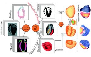
Specific developments include:
- Automated analysis (segmentation, registration) of in vivo and in vitro cardiac images
- Specific methods for the quantification of cardiac Diffusion MRI scans
- Generation of volume meshes from images
- Study, using cardiac models, of the effect on electrical and mechanical effects of normal myocardial structure and their disruption in pathologies such as myocardial infarction and hypertrophic cardiomyopathy.
This is done in collaboration with the teams of Prof. Peter Kohl at the National Lung and Heart Institute, Dr. Jurgen Schneider at the Wellcome Centre for Human Genetics, and Dr. Erica Dall'Armellina at the Oxford Acute Vascular Imaging Centre (AVIC).
 Newt
Newt